


 Latinoamérica
 Europa
Multicentric Lymphoma in a Sheep









The lymphoma in sheep is an uncommon pathology with nonspecific symptoms that can resemble other diseases, with the enlargement of lymph nodes in different regions of the body being a suggestive finding of this pathology. Pathological anatomy and immunohistochemistry are essential for a definitive diagnosis, but, in the absence of treatment, euthanasia is the most appropriate option, both from an economic standpoint and for the animal’s welfare.
 Below is a clinical case of an adult Lacaune sheep referred to the Ruminant Clinical Service at the Veterinary Faculty of Zaragoza with severe respiratory symptoms, whose study led to the definitive diagnosis. Below is a clinical case of an adult Lacaune sheep referred to the Ruminant Clinical Service at the Veterinary Faculty of Zaragoza with severe respiratory symptoms, whose study led to the definitive diagnosis. |
ANAMNESIS: INDICATIONS OF A DISEASE IN THE SHADOW
In October 2023, the Animal Experimentation Service (SEA) of the University of Zaragoza referred to the Ruminant Clinical Service (SCRUM) of the Veterinary Faculty of Zaragoza a six-year-old adult Lacaune sheep, of dairy aptitude.
 The animal exhibited evident respiratory symptoms that were life-threatening. The sheep was permanently housed in a lot, under intensive regime, within the SEA facilities at the Faculty of Veterinary Medicine.
 Suspecting a pneumonic condition, treatment was initiated. However, the clinical deterioration and the severity presented by the animal necessitated its referral for a more comprehensive study. Suspecting a pneumonic condition, treatment was initiated. However, the clinical deterioration and the severity presented by the animal necessitated its referral for a more comprehensive study. |

CLINICAL SIGNS: CLUES THAT TAKE YOUR BREATH AWAY
Upon arrival, the animal underwent a thorough clinical examination.
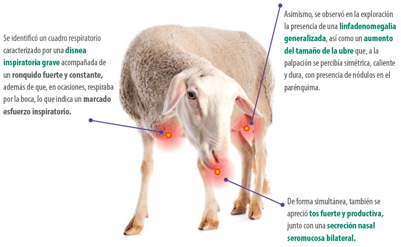
DIAGNOSIS: CONNECTING THE DOTS IN CLINICAL RESEARCH
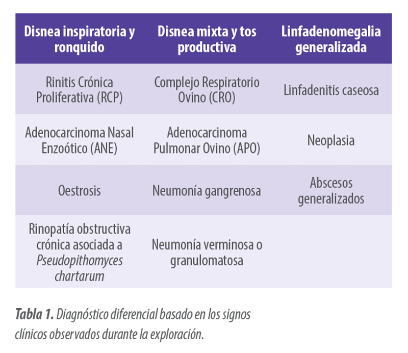
Differential Diagnosis
1. Pathologies affecting the upper respiratory tract
The symptoms presented by the animal, especially the marked inspiratory dyspnea, directed the diagnosis towards an upper respiratory tract pathology. Based on this suspicion, several pathologies were included in the differential:
 Chronic Proliferative Rhinitis (CPR), caused by Salmonella enterica subsp. diarizonae serovar 61:k:1,5,(7) (Meehan et al., 1992).
This individual pathology causes a progressive inflammation of the ventral nasal turbinates, characterized by inspiratory dyspnea, snoring, and dense mucous nasal discharge that can be unilateral or bilateral. Occasionally, enlarged regional lymph nodes may be observed.
Based on experience and clinical signs, it seemed the most feasible option and, due to the severity of the condition, a sample was taken and the animal was treated for it with anti-inflammatories and marbofloxacin, the treatment of choice against this agent (Rubira et al., 2019).
Oestrosis, cavitary myiasis caused by the larvae of the Oestrus ovis fly, considered the most important and prevalent rhinitis in warm and dry geographical areas (Gracia et al., 2010).
It is usually associated with rearing in extensive or grazing conditions, being a collective, herd pathology (Lucientes et al., 1998). Animals present mucous or purulent nasal discharge, even hemorrhagic (Gracia et al., 2010).
Enzootic Nasal Adenocarcinoma (ENA), contagious tumor caused by a betaretrovirus known as ENTV (Enzootic Nasal Tumor Virus) that affects sheep (ENTV-1) and goats (ENTV-2).
This pathology causes a neoplastic transformation of the secretory epithelial cells of the ethmoidal turbinate, leading to inspiratory dyspnea, continuous serous nasal discharge, and even exophthalmos and cranial deformation due to tumor growth (De las Heras et al., 2003; Walsh et al., 2013).
Obstructive rhinopathy associated with the fungus Pseudopithomyces chartarum, a collective pathology characterized by inspiratory dyspnea caused by a chronic nasal obstruction, total or partial, at the level of the nostrils, in addition to a hepatopathy (De las Heras et al., 2022).
2. Pathologies affecting the lower respiratory tract
Simultaneously, the strong and productive cough also seemed to indicate involvement in the lower respiratory tract, so the differential also included other pathologies such as:
 Ovine Respiratory Complex (ORC), caused by bacteria such as Mannheimia haemolytica, Pasteurella multocida, Bibersteinia trehalosi and Mycoplasma spp that act opportunistically (Lacasta et al., 2019).
Ovine Pulmonary Adenocarcinoma (OPA), a lung tumor caused by the Jaagsiekte sheep retrovirus (JSRV), a member of the Retroviridae family (De las Heras et al., 2005; Griffiths et al., 2010) that leads to severe conditions in advanced stages, with nasal discharge and severe respiratory distress.
Gangrenous pneumonia, generally caused by the inhalation of foreign bodies and subsequent reaction when these lodge in the lung (Lacasta et al., 2019).
Verminous pneumonia or granulomatous pneumonia, a disease caused by lung nematodes, although it is more typical of non-intensive production systems.
3. Pathologies causing generalized lymphadenomegaly
The animal presented with generalized lymphadenomegaly, which could suggest the presence of systemic processes, such as:
Generalized infection or abscesses.
Neoplasms, such as multicentric lymphoma, characterized by the enlargement of lymph nodes (Johnstone and Manktelow, 1978).
Superficial caseous lymphadenitis, caused by the bacterium Corynebacterium pseudotuberculosis.
 Lymphadenomegaly was also observed at the udder level, which, together with the findings in the palpation of the udder and the animal’s dairy aptitude, did not allow the exclusion of mastitis of bacterial etiology (Smith et al., 2015).
Complementary tests performed
A hematology was performed, which showed microcytic and hypochromic anemia accompanied by neutrophilia, eosinopenia, lymphopenia, and thrombocytopenia, suggestive of a chronic inflammatory or neoplastic process.
A thermographic study was also conducted due to the diagnostic value of this test in diagnosing upper airway respiratory processes (Ferrer et al., 2021).
 Thermography revealed the bilateral presence of areas with increased temperature at the level of the nasal cavities, both in frontal and lateral views, suggestive of inflammation.
 Due to the clinical signs and the results of complementary tests, especially thermography, it was decided to perform a nasal sampling to confirm or rule out the presence of an RCP condition, the main suspicion. Due to the clinical signs and the results of complementary tests, especially thermography, it was decided to perform a nasal sampling to confirm or rule out the presence of an RCP condition, the main suspicion.
|
 The negative results obtained in PCR against its causal agent ruled out this disease, which is why the antibiotic treatment was stopped, maintaining the anti-inflammatory due to the clinical improvement the animal showed after its application.
The animal eventually died due to the severity of the clinical condition it presented, which led to a thorough postmortem study by the Department of Animal Pathology at the University of Zaragoza.
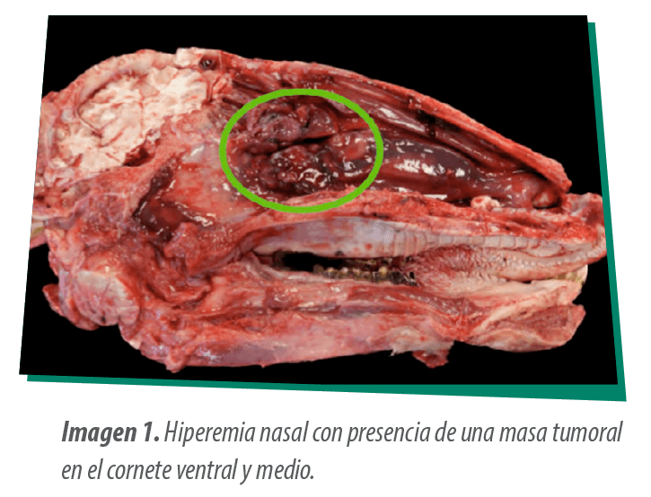
A generalized nasal hyperemia was observed accompanied by a tumor mass about 6 cm in diameter affecting the nasal mucosa of the caudal half of the ventral turbinate and the middle turbinate (Image 1).
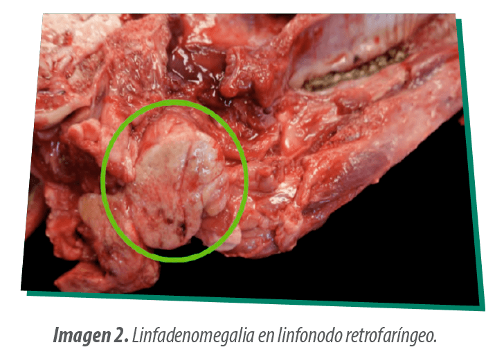
Another mass of similar appearance was observed in the retropharyngeal lymph node which presented lymphadenomegaly, with a shiny whitish color (Image 2).
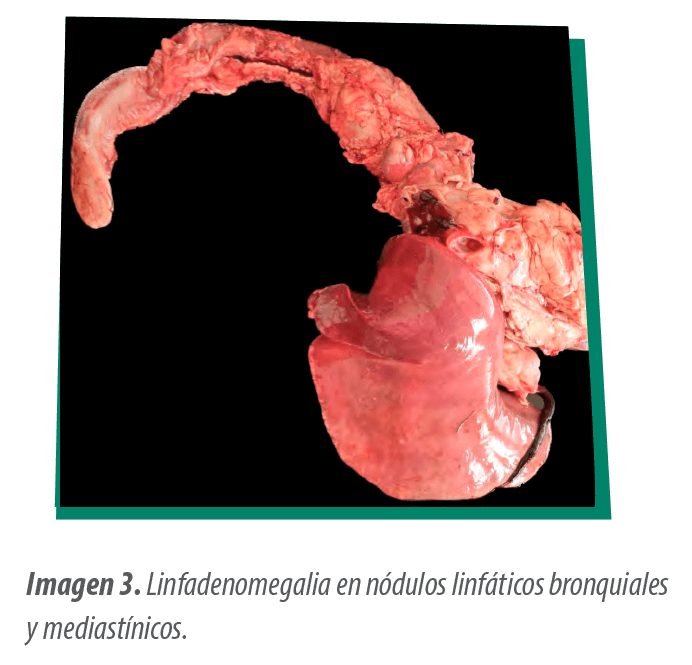
In the thoracic cavity, the bronchial and mediastinal lymph nodes showed a similar appearing lymphadenomegaly surrounding the trachea (Image 3).
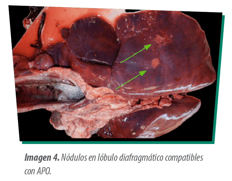
On the dorsal surface of the right diaphragmatic lobe of the lung, hard and whitish nodulations about 2 cm in size were also observed, concentrically surrounded by other similar ones but smaller (<0.5 cm) (Image 4). These nodulations deepened on sectioning, being macroscopically compatible with an APO or with satellite nodules of the masses observed in the different lymph nodes.
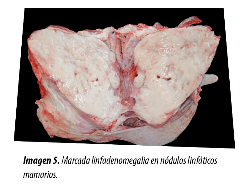
At the udder level, the mammary lymph nodes showed marked lymphadenomegaly (approximately 25×15 cm) with a shiny whitish appearance on sectioning (Image 5), although the mammary gland did not show apparent lesions.
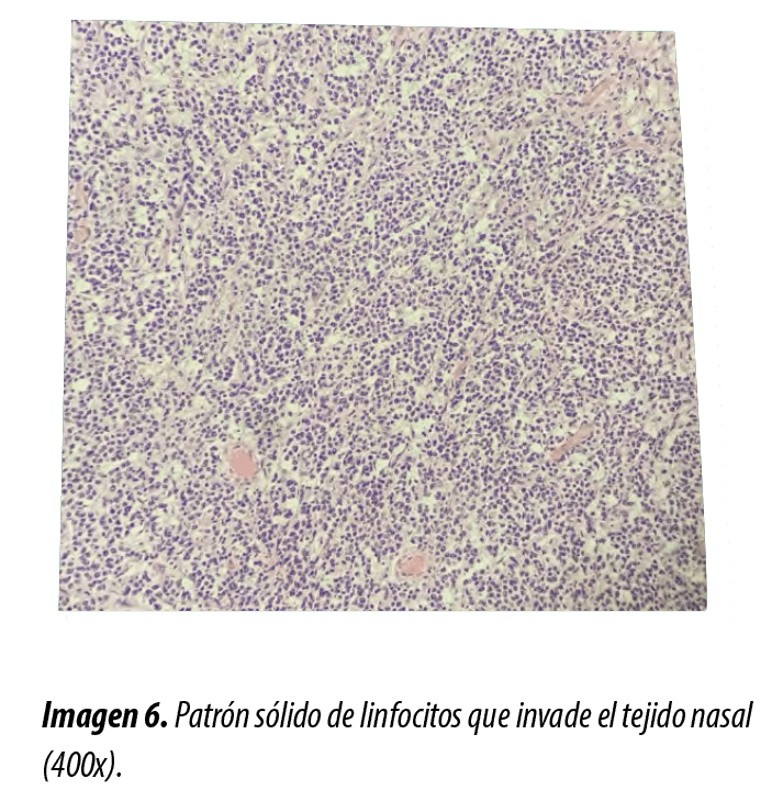
The histopathology of the masses at the nasal level and in the lymph nodes revealed that they were composed of a large number of tumor-like cells, compatible with neoplastic lymphocytes in a solid pattern that profoundly altered the normal tissue architecture, being an aspect compatible with a lymphoma (Image 6).
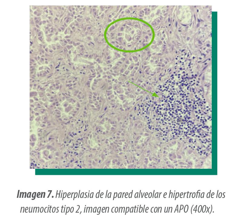
In contrast, the nodules found in the diaphragmatic lobe of the lung corresponded to an increase in the size of the type 2 pneumocytes, indicating a hyperplasia of the alveolar wall. Additionally, fibrosis was observed, a notable inflammatory infiltrate composed of macrophages and other cells, as well as areas of necrosis (Image 7), a characteristic pattern of OPD.
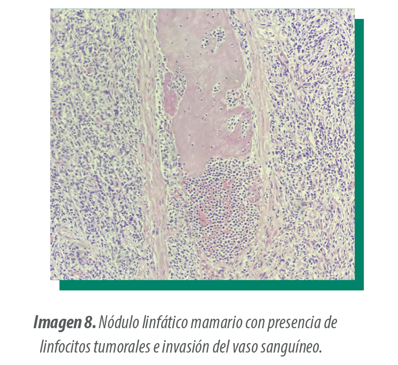
The mammary lymph node also showed the presence of tumor lymphocytes that caused the destruction of organic territories and its enlargement denoting organ hyperplasia (Image 8). On the other hand, the mammary gland also showed an invasion of neoplastic lymphocytes despite its apparently healthy macroscopic appearance.
PRESUMPTIVE DIAGNOSIS: RECONSTRUCTING THE CLINICAL PUZZLE
The final diagnosis included the presence of two types of neoplasia simultaneously, an APO and a multicentric lymphoma.
Immunohistochemical tests were performed to determine the predominant lymphocyte series in the lymphoma so that, due to its reaction with the Pax5 biomarker, present in the membranes of B lymphocytes, it was considered as a type B multicentric lymphoma.
PATHOGENESIS OF MULTICENTRIC LYMPHOMA
Multicentric lymphoma is a common disease in domestic species such as dogs or dairy cattle. Even in goats, some sporadic cases have been described. However, very few cases have been described in sheep, so it is considered a rare disease.
In small ruminants, clinical signs are nonspecific and include progressive weight loss, cachexia, apathy, fever, and dyspnea (Digrassie et al., 1997; Anjos et al., 2010; Rego et al., 2015), with diagnosis occurring post-mortem and accidentally.
 However, biopsies of peritoneal fluid or enlarged lymph nodes, where a high number of immature and degenerated lymphocytes can be observed, may be useful for the in vivo diagnosis of this type of disease (Santos et al., 2020).
Regarding the progression of tumors, a slaughterhouse study conducted by Jonhstone and Manktelow (1978), which analyzed the location of tumors in 22 sheep with malignant lymphoma, found that the most consistent form of this lymphoma is the enlargement of the affected lymph nodes, followed by the progressive spread of neoplastic tissue to other lymph nodes and non-lymphoid organs, as seen in this case in the udder and nasal turbinate.
After the lymph nodes, the spleen is the second most affected organ, followed by its presence in the uterus. However, it is important to note that the spread to other organs as a result of systemic lymphoma is very common, which leads to a very unfavorable prognosis.
| According to the WHO criteria, adapted to specific species (Valli et al., 2002), the histopathological classification of a lymphoma is based on:
|
 |
Kiser and Löhr (2017) adapted these criteria to the goat species, classifying according to:
In sheep, these values might be slightly altered, but can be used. |
 Topographic distribution.
 Distribution (diffuse or nodular).
Regarding the cause of the tumor, most cases are spontaneous, with a clear genetic and immunogenic component (Jonhstone and Manktelow, 1978).
However, there are several studies that link the bovine leukemia virus (BLV) with the occurrence of malignant multicentric lymphoma in sheep (Murakami, 1994; Kenyon, 1981). However, Spain is free of BLV. For this reason, a specific inducing cause of the appearance of the neoplasia in this case is unknown.
Continuing with the diagnosis of the animal, it is important to note that, in addition to the multicentric lymphoma, hard nodules were located in the right diaphragmatic lobe of the lung that deepened on section, diagnosed as APO, a tumor of viral etiology frequently observed in sheep.
| The confluence of these two pathologies is noteworthy, not only because of the low probability of an animal presenting two simultaneous neoplastic processes, but because Di guardo et al. (1992) described a case with similar characteristics, where APO was observed alongside multicentric lymphoma in an adult sheep of two years. Therefore, although it could be a coincidence, there might be a possibility that there could be some relationship between both pathologies. |
TREATMENT AND PREVENTION: CLOSING THE LOOP
Treatment for this type of pathologies is unfeasible at a commercial level in sheep or small ruminants. In other domestic species, such as dogs, surgery and chemotherapeutic treatments are therapeutic options used (Toyoda et al., 2023). Even, rabacfosamide has shown good results in this species (Imveterinaria 2021).
Currently, it is not possible to apply these treatments in livestock, so the slaughter of the animal remains the most appropriate measure to prevent clinical progression and deterioration of the animal.
It is not possible to apply preventive measures against these tumors, as, like most neoplasms, their occurrence is influenced by genetic and environmental characteristics, which prevents establishing a preventive pathway.
| This case highlights the diagnostic complexity of multicentric lymphoma in sheep and the need for a detailed clinical and pathological approach. Although there are no viable therapeutic options in the species, its study provides valuable information about a rare pathology, helping to improve its recognition and management in veterinary practice. |
BIBLIOGRAPHY
Anjos, B.L.; Trost, M.E.; Diefenbach, A.; Gressler, L.T.; Irigoyen, L.F., 2010. Linfossarcoma de células B multicêntrico em uma ovelha. Acta Scientiae Veterinariae, v.38, n.3, p.315-318.
De las Heras, M., Ortin, A., Cousens, C., Minguijon, E., Sharp, J.M., 2003. Enzootic nasal adenocarcinoma of sheep and goats. In: In: Fan, H. (Ed.), Jaagsiekte Sheep Retrovirus and Lung Cancer. Curr. Top. Microbiol. 275, 201–223.
De las Heras, M., Ortín, A., Salvatori, D., de Villareal, M. P., Cousens, C., et al., 2005. A PCR technique for the detection of Jaagsiekte sheep retrovirus in the blood suitable for the screening of ovine pulmonary adenocarcinoma in field conditions. Research in veterinary science, 79(3), 259-264.
De las Heras, M., Lacasta, D., Reséndiz, R. A., Rivas, A., Garzianda, A., de Miguel, R., … & Ferrer, L. M.. 2022. Chronic pithomycotoxicosis associated with obstructive rhinopathy in sheep. Veterinary Pathology, 59(6), 950-959.
Digrassie, W.A.; Wallace, M.A.; Sponenberg, D.P., 1997. Multicentric lymphosarcoma with ovarian involvement in a Nubian goat. The Canadian veterinary journal, v.38, n.6, p.383-384.
Di Guardo, G., Condoleo, R., & Autorino, G. L., 1992. Multicentric lymphosarcoma associated with pulmonary adenomatosis (Jaagsiekte), pulmonary lymphoid hyperplasia, and lymphoid interstitial pneumonia in a ewe. Veterinary pathology, 29(3), 262–264.
Ferrer, L.M., Ramos, J.J., Castells, E., Ruiz, H., Climent M. & Lacasta, D., 2021. Use of computed tomography and thermography for the diagnosis of respiratory disorders in adult sheep.
Gracia, M.J., Lucientes, J., Peribáñez, M.A., Castillo, J.A., Calvete, C., Ferrer, L.M., 2010. Epidemiology of Oestrus ovis infection of sheep in Northeast Spain (mid-Ebro Valley). Trop. Anim. Health Pro. 42, 811–813.
Griffiths, D. J., Martineau, H. M., & Cousens, C., 2010. Pathology and pathogenesis of ovine pulmonary adenocarcinoma. Journal of comparative pathology, 142(4), 260-283. 29.
Imveterinaria. Rescue therapies for the treatment of multicentric lymphoma in dogs. 2021. Johnstone, A. C., & Manktelow, B. W., 1978. The pathology of spontaneously occurring malignant lymphoma in sheep. Veterinary pathology, 15(3), 301–312.
Kenyon, S.J.; Ferrer, J.F.; McFleey, R.A.; Graves, D.C., 1981. Induction of lymphosarcoma in sheep by bovine leukemia virus. Journal of the National Cancer Institute, v.67, n.5, p.1157-1163.
Kiser, P. K., & Löhr, C. V. (2017). Lymphoma Classification in Goats. Veterinary pathology, 54(4), 611 619.
Lacasta D., Fernández A., González J.M., Ramos J.J., Ortín A., Ferrer L.M., 2019. Gangrenous pneumonia, ovine respiratory complex and visceral form of T caseous lymphadenitis: Relevance in lower respiratory tract disorders of adult sheep. Small Ruminant Research. 180, 100-105.
Lucientes, J., Castillo, J.A., Ferrer, L.M., Peribáñez, M., Ferrer Dufol, M., Gracia-Salinas, M.J., 1998. Efficacy of orally administered ivermectin against larval stages of Oestrus ovis in sheep. Vet. Parasitol. 75, 255–259.
Meehan, J.T., Brogden, K.A., Courtney, C., Cutlip, R.C., Lehmkuhl, H.D., 1992. Chronic proliferative rhinitis associated with Salmonella arizonae in sheep. Vet. Pathol. 29, 556–559.
Murakami, K.; Okada, K.; Irawa, Y.; Aida, Y., 1994. Bovine leukemia virus induces CD5 – Bcells lymphoma in sheep despiste temporarily increasing CD5 + Bcells in asyntomatic stage. Virology, v.202, p. 458-465.
Rego, O.; Souza, J.C.A.; Mendonça, C.L.; Silva, R.J.; Silva, N.A.A.; Riet-Correa, F.; Afonso, J.A.B., 2015. Spontaneous occurrence of malignant lymphoma in a sheep: case report. Ciência Veterinária nos Trópicos, Recife-PE, v.18, n.1, p.26-31.
Rubira, I., Figueras, L., De las Heras, M., Bueso, J. P., Castells, E., Climent, M., & Lacasta, D., 2019. Chronic proliferative rhinitis in sheep: An update. Small Ruminant Research, 179, 21-25.
Smith, E. M., Willis, Z. N., Blakeley, M., Lovatt, F., Purdy, K. J., & Green, L. E., 2015. Bacterial species and their associations with acute and chronic mastitis in suckler ewes. Journal of dairy science, 98(10), 7025–7033.
Santos, E., Kalb, A. L., Barbosa, A. A., Rabassa, V. R., Correa, M. N., 2020. Multicentric lymphoma in Sheep (case report), Revista Brasileira de Saúde e Produção Animal, 21.
Toyoda, H., Tani, A., Goto‐Koshino, Y., Motegi, T., Sakamoto, M., Mochizuki, T., Harada, K., Kobayashi, T., Setoguchi, A., Shizuta, Y., Mizuno, T., Irie, M., Nakamichi, J., Tsujimoto, H., Ohmi, A., Fukuoka, R., Nakamura, Y. & Tomiyasu, H., 2023. Gene expression profiles associated with early relapse during first remission induction in canine multicentric high-grade B-cell lymphoma. The Journal of veterinary medical science, 86(1), 18-27.
Valli VE, Jacobs RM, Parodi AL, Vernau W, Moore PF., 2002. Histological Classification of Hematopoietic Tumors of Domestic Animals.
World Health Organization International Classification of Tumors in Domestic Animals. Washington, DC: Armed Forced Institute of Pathology.
Walsh, S.R., Linnerth-Petrik, N.M., Yu, D.L., Foster, R.A., Menzies, P.I., Diaz-Méndez, A., Wootton, S.K., 2013. Experimental transmission of enzootic nasal adenocarcinoma in sheep. Vet. Res. 44 (1), 66.
Anjos, B.L.; Trost, M.E.; Diefenbach, A.; Gressler, L.T.; Irigoyen, L.F., 2010. Multicentric B-cell lymphosarcoma in a sheep. Acta Scientiae Veterinariae, v.38, n.3, p.315-318.
De las Heras, M., Ortin, A., Cousens, C., Minguijon, E., Sharp, J.M., 2003. Enzootic nasal adenocarcinoma of sheep and goats. In: In: Fan, H. (Ed.), Jaagsiekte Sheep Retrovirus and Lung Cancer. Curr. Top. Microbiol. 275, 201–223.
De las Heras, M., Ortín, A., Salvatori, D., de Villareal, M. P., Cousens, C., et al., 2005. A PCR technique for the detection of Jaagsiekte sheep retrovirus in the blood suitable for the screening of ovine pulmonary adenocarcinoma in field conditions. Research in veterinary science, 79(3), 259-264.
De las Heras, M., Lacasta, D., Reséndiz, R. A., Rivas, A., Garzianda, A., de Miguel, R., … & Ferrer, L. M.. 2022. Chronic pithomycotoxicosis associated with obstructive rhinopathy in sheep. Veterinary Pathology, 59(6), 950-959.
Digrassie, W.A.; Wallace, M.A.; Sponenberg, D.P., 1997. Multicentric lymphosarcoma with ovarian involvement in a Nubian goat. The Canadian veterinary journal, v.38, n.6, p.383-384.
Di Guardo, G., Condoleo, R., & Autorino, G. L., 1992. Multicentric lymphosarcoma associated with pulmonary adenomatosis (Jaagsiekte), pulmonary lymphoid hyperplasia, and lymphoid interstitial pneumonia in a ewe. Veterinary pathology, 29(3), 262–264.
Ferrer, L.M., Ramos, J.J., Castells, E., Ruiz, H., Climent M. & Lacasta, D., 2021. Use of computed tomography and thermography for the diagnosis of respiratory disorders in adult sheep.
Gracia, M.J., Lucientes, J., Peribáñez, M.A., Castillo, J.A., Calvete, C., Ferrer, L.M., 2010. Epidemiology of Oestrus ovis infection of sheep in Northeast Spain (mid-Ebro Valley). Trop. Anim. Health Pro. 42, 811–813.
Griffiths, D. J., Martineau, H. M., & Cousens, C., 2010. Pathology and pathogenesis of ovine pulmonary adenocarcinoma. Journal of comparative pathology, 142(4), 260-283. 29.
Imveterinaria. Rescue therapies for the treatment of multicentric lymphoma in dogs. 2021.
Johnstone, A. C., & Manktelow, B. W., 1978. The pathology of spontaneously occurring malignant lymphoma in sheep. Veterinary pathology, 15(3), 301–312.
Kenyon, S.J.; Ferrer, J.F.; McFleey, R.A.; Graves, D.C., 1981. Induction of lymphosarcoma in sheep by bovine leukemia virus. Journal of the National Cancer Institute, v.67, n.5, p.1157-1163.
Kiser, P. K., & Löhr, C. V. (2017). Lymphoma Classification in Goats. Veterinary pathology, 54(4), 611–619.
Lacasta D., Fernández A., González J.M., Ramos J.J., Ortín A., Ferrer L.M., 2019. Gangrenous pneumonia, ovine respiratory complex and visceral form of T caseous lymphadenitis: Relevance in lower respiratory tract disorders of adult sheep. Small Ruminant Research. 180, 100-105.
Lucientes, J., Castillo, J.A., Ferrer, L.M., Peribáñez, M., Ferrer Dufol, M., Gracia-Salinas, M.J., 1998. Efficacy of orally administered ivermectin against larval stages of Oestrus ovis in sheep. Vet. Parasitol. 75, 255–259.
Meehan, J.T., Brogden, K.A., Courtney, C., Cutlip, R.C., Lehmkuhl, H.D., 1992. Chronic proliferative rhinitis associated with Salmonella arizonae in sheep. Vet. Pathol. 29, 556–559.
Murakami, K.; Okada, K.; Irawa, Y.; Aida, Y., 1994. Bovine leukemia virus induces CD5 – Bcells lymphoma in sheep despiste temporarily increasing CD5 + Bcells in asyntomatic stage. Virology, v.202, p. 458-465.
Rego, O.; Souza, J.C.A.; Mendonça, C.L.; Silva, R.J.; Silva, N.A.A.; Riet-Correa, F.; Afonso, J.A.B., 2015. Spontaneous occurrence of malignant lymphoma in a sheep: case report. Ciência Veterinária nos Trópicos, Recife-PE, v.18, n.1, p.26-31.
Rubira, I., Figueras, L., De las Heras, M., Bueso, J. P., Castells, E., Climent, M., & Lacasta, D., 2019. Chronic proliferative rhinitis in sheep: An update. Small Ruminant Research, 179, 21-25.
Smith, E. M., Willis, Z. N., Blakeley, M., Lovatt, F., Purdy, K. J., & Green, L. E., 2015. Bacterial species and their associations with acute and chronic mastitis in suckler ewes. Journal of dairy science, 98(10), 7025–7033.
Santos, E., Kalb, A. L., Barbosa, A. A., Rabassa, V. R., Correa, M. N., 2020. Multicentric lymphoma in Sheep (case report), Revista Brasileira de Saúde e Produção Animal, 21.
Toyoda, H., Tani, A., Goto‐Koshino, Y., Motegi, T., Sakamoto, M., Mochizuki, T., Harada, K., Kobayashi, T., Setoguchi, A., Shizuta, Y., Mizuno, T., Irie, M., Nakamichi, J., Tsujimoto, H., Ohmi, A., Fukuoka, R., Nakamura, Y. & Tomiyasu, H., 2023. Gene expression profiles associated with early relapse during first remission induction in canine multicentric high-grade B-cell lymphoma. The Journal of veterinary medical science, 86(1), 18-27.
Valli VE, Jacobs RM, Parodi AL, Vernau W, Moore PF., 2002. Histological Classification of Hematopoietic Tumors of Domestic Animals. World Health Organization International Classification of Tumors in Domestic Animals. Washington, DC: Armed Forced Institute of Pathology.
Walsh, S.R., Linnerth-Petrik, N.M., Yu, D.L., Foster, R.A., Menzies, P.I., Diaz-Méndez, A., Wootton, S.K., 2013. Experimental transmission of enzootic nasal adenocarcinoma in sheep. Vet. Res. 44 (1), 66.