


 Latinoamérica
 Europa
Contagious Nodular Dermatosis: Key Strategies to Halt the Spread of This Dangerous Disease


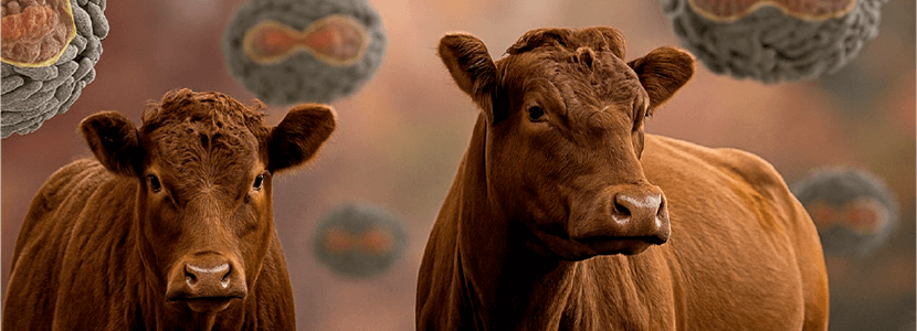











In this era we are living in, emerging diseases that affect both humans and different animal species—or both at the same time—are taking on a prominence that means that almost every year, we have to face one or several health crises simultaneously.
This year, unfortunately, we have been hit by one of the big ones, one of the “ugliest”: contagious nodular dermatosis.
 It is a viral infection caused by a virus belonging to the genus Capripoxvirus, representative of the viruses encompassed in the classic “poxes,” which approximately a decade ago had made a significant incursion in Greece and the Balkan countries.
Never before had it positioned so close to us, to the point that, finally, it has fully affected us.
 Just before the summer, its presence in Italy and, above all, in France did not bode well for our interests.
 Unfortunately, in the month of October all our fears were confirmed and the infection has appeared in Spanish territory, specifically in the province of Girona.
| Despite the difficulty and complexity of the infection, there are some facts that should make us at least optimistic about the possibilities of eliminating such a problematic enemy. It won’t be easy, but we have some tools for the fight that we do not have against other infections.
|

FROM AFRICA TO THE MEDITERRANEAN: THE JOURNEY OF THE VIRUS
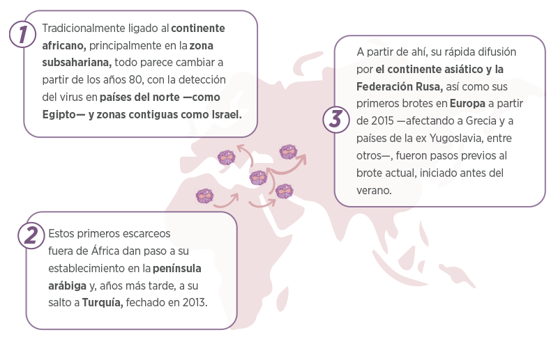
| It should be noted that, in those clinical events, occurring mainly between 2015 and 2016, the number of affected groups exceeded 7,500. In contrast, currently, the number of affected groups in Europe is much lower —not reaching 200—, although, logically, it continues to increase. |
THE VIRUS – A CLOSE RELATIVE OF SHEEP AND GOAT POX
The causative agent of contagious nodular dermatosis is a Capripoxvirus, genetically very similar to the sheep and goat pox viruses.
 The latter, in its sheep version, was detected just a few years ago in certain areas of Andalusia and Castilla-La Mancha.
Both viruses are so close that, from a serological point of view, they are indistinguishable, which demonstrates their close antigenic similarity.
 Even so, they exhibit a marked host specificity: while nodular dermatitis primarily affects cattle, sheep and goats are not considered species with a relevant epidemiological role in this infection.
The virus is not particularly resistant to common disinfectants, although, when it is shed from the animal in the typical scabs that form after several weeks of clinical progression, it can survive for prolonged periods, even close to a month.
| This aspect is key when designing disease control and eradication programs. |
SUSCEPTIBLE SPECIES AND EPIDEMIOLOGICAL ROLE
Cattle, main host
The cattle —especially Bos taurus— constitute the target species of this infectious agent. However, zebu (Bos taurus indicus) and even water buffalo (Bubalus bubalis) are also susceptible to infection, although to a lesser extent.
 In the first case, the maximum average morbidity is around 30%, a value that is significantly reduced in its African and Asian relatives.
As the infection has spread to new geographical areas, it has been observed that other species, such as yaks, are also susceptible to infection.
| In cattle, all breeds and animals of any age can be affected, although young animals seem to be more sensitive to the infection, making it important to consider the existence of a significant “individual factor” in susceptibility to the infectious agent. |
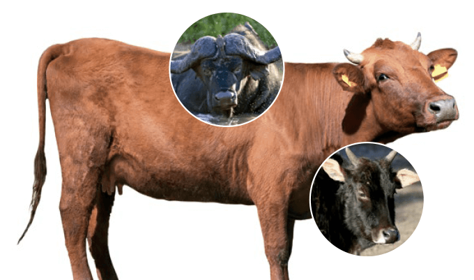
 Thus, within the same group, individuals with evident symptoms can be observed living alongside others who, despite being infected and in the viremic phase, do not develop clinical signs.
| Contagious nodular dermatosis is not considered a zoonosis, it does not affect humans, either directly or indirectly. |
Wildlife: an open question
It is unknown whether wildlife plays any epidemiological role in maintaining the virus. However, in Africa—where the disease originates—it has been demonstrated directly or indirectly in various species, such as impalas or kudus, and clinical cases compatible with the disease have been observed in others, such as springboks and even camelids.

| For now, it has not been determined if any European species could be susceptible to the virus and contribute to its maintenance or transmission. |
HOW CONTAGIOUS NODULAR DERMATOSIS SPREADS
Short-distance transmission
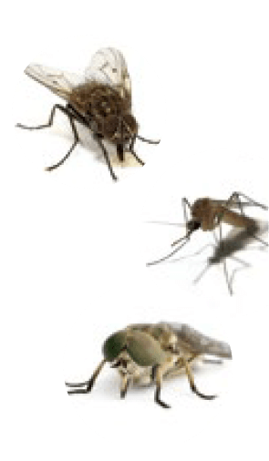 At short distances, the transmission of the agent between groups is mainly associated with the action of vectors (flies, mosquitoes, horseflies, among others), although not many species have been consistently identified with the spread of the infection.
 It is a mechanical transmission in which the virus adheres to the legs or other parts of the vector, allowing the contaminated insect to transmit the agent to other hosts for a period of 5-6 days.
 It is estimated that the infection could advance 1-2 km daily through this mechanism, although, obviously, this value depends on numerous factors.
| Since vectors play an essential role, the infection mainly manifests during the times of the year when they are most active. However, with the current conditions of climate change and rising temperatures, these estimates should be interpreted with caution. |
Direct transmission of the virus at short distances cannot be ruled out, as infected animals shed it for several weeks (3 to 4 weeks) through various secretions and excretions (oral, nasal, conjunctival, milk, etc.).
 However, direct contact between hosts does not seem to be a particularly effective means of contagion, to the point of not justifying on its own the transmission patterns observed in the field.
| The possibility of foodborne, iatrogenic, or other transmission from contaminated material cannot be excluded either, although it does not seem to have sufficient capacity to keep the infection active in an area. Finally, the available data on congenital transmission are scarce and do not firmly support this hypothesis. |
In Africa, some species of ticks also seem to play an important role in the transmission of the infection.
In this case, we could talk about biological transmission between the different stages of these vectors, which could facilitate the survival of the virus during less favorable times of the year, such as winter and other periods with fewer vectors present.
Long-distance transmission
Over long distances, transmission seems more linked to the movement of individuals or contaminated material, which would explain the spread of the infection over many kilometers, a phenomenon difficult to attribute solely to the role of vectors. Therefore, it is essential to maintain a strict control of movements in affected areas.
HOW THE DISEASE MANIFESTS IN LIVESTOCK
In affected herds, where morbidity is variable, the infection is characterized by a biphasic febrile period, which may be accompanied by nasal, oral, or ocular involvement and secretions (in the latter case, potentially causing ulcerations and corneal opacity).
 The presence of skin lesions in different areas of the body, both external and internal, is common.
The most characteristic feature is the appearance of cutaneous nodules which, although in some animals are limited to regions such as the head, muzzle, or legs, in many cases are distributed generally, also affecting the mammary gland or genitals.
These externally visible lesions can also be found internally, especially in the oral cavity and trachea, as well as in other locations of the respiratory and digestive systems. In these areas, congestion and hemorrhage are common.
Both externally and internally, the lesions are accompanied by inflammatory processes that affect, for example, lymphoid tissues, which increase significantly in size. The inflammation also causes edema in anatomical regions such as the chest and limbs, which can lead to lameness.
 The condition evolves over several weeks, with necrosis of the external area of the nodules, which, like an inverted cone, penetrates the skin, sometimes reaching the muscle tissue.
 The mortality is variable and, although outbreaks with high percentages have been described, it rarely exceeds 2–3% of the affected animals.
The immune response is detected several weeks after the onset of the clinical condition, and in animals that overcome the infection, the cutaneous lesions may remain visible up to a year after the clinical episode.
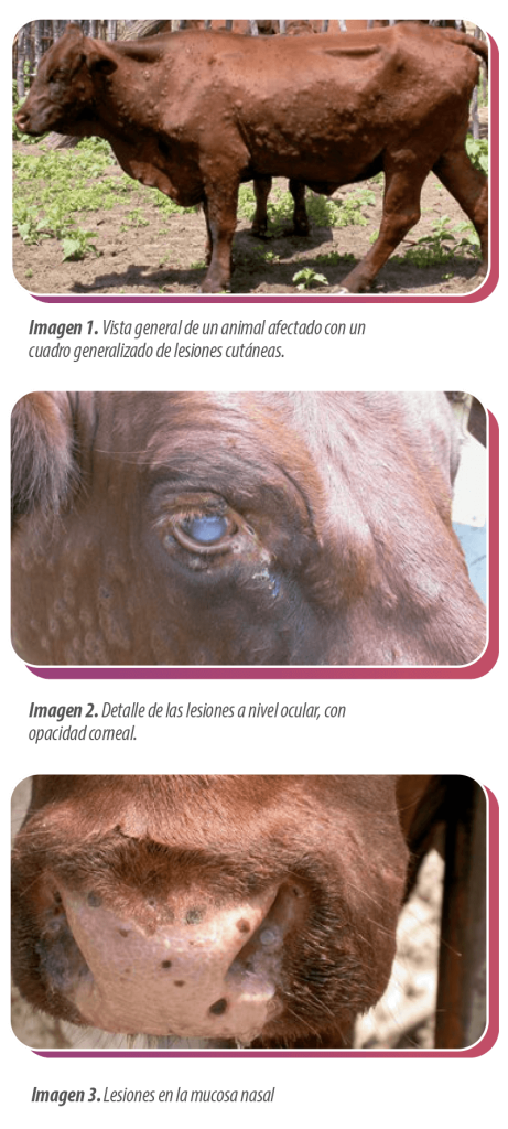
Images courtesy of Prof. M. Scacchia, Istituto Zooprofilattico Sperimentale dell´Abruzzo e del Molise “G. Caporale”, Italy

CONTROL AND ERADICATION STRATEGIES
Immediate containment measures
As it is an infection included in the European Union list A, its suspicion must immediately trigger the implementation of urgent eradication strategies.
A sanitary clearance of all animals in the affected group must be carried out, as well as the destruction of carcasses and treatment of contaminated material.
The delineation of protection and surveillance zones (20 and 50 km around the outbreaks, respectively) entails strict movement control, authorized only in certain cases, such as the transport of animals to slaughterhouses.
The milk from restricted areas can be transported for heat treatment, provided the transport trucks are sealed and hermetically closed.
Emergency vaccination: the key tool
These measures are complemented by the use of live homologous vaccines against this virus (produced with the bovine virus), which are administered “in emergency” to animals located within a 50 km radius around clinical outbreaks.
 Based on previous experience from the 2015–2017 period, these vaccines have proven effective in containing and eradicating the disease.
In this regard, it is essential to act quickly to:
1 Achieve a high vaccination coverage as soon as possible.
2 Immediately report any suspected new outbreaks.
 The European Union’s action protocol is based on the experience gained during the outbreaks recorded in Europe a decade ago, when the infection was finally eradicated after several years of work. The European Union’s action protocol is based on the experience gained during the outbreaks recorded in Europe a decade ago, when the infection was finally eradicated after several years of work. |
 It is essential to vaccinate all susceptible animals near affected areas as soon as possible in order to contain its spread. All of this must be accompanied by:
 Strict movement control measures.
Treatment of contaminated material.
Rapid diagnosis in all those farms where the presence of the virus is suspected.
Diagnosis and differential diseases
In these farms, the diagnosis is fundamentally direct, through PCR performed from the skin lesions of affected animals or swab samples collected from individuals with symptoms compatible with the infection. In the case of contagious nodular dermatosis, we have tools that allow us to move towards its eradication.
 Although with various prognoses and presentations, other skin diseases must be ruled out such as dermatophilosis, besnoitiosis, dermatophytosis or papillomatosis, as well as other non-contagious processes, including episodes of photosensitization.
Eradication: a fight that requires the involvement of everyone
In the case of contagious nodular dermatosis, we have tools that allow us to move towards its eradication.
This disease, in addition to the direct damage it causes to the affected herds, has a considerable economic and commercial impact due to the movement restrictions imposed at the national level. Therefore, the involvement of the entire livestock and technical sector is essential to achieve infection control as soon as possible.
| We will see in the coming months the evolution of this struggle, which will not be easy or short, but which we hope will culminate successfully. |

You may be interested: Ovine and Caprine Pox – The resurgence of an old acquaintance