


 Latinoamérica
 Europa
First Clinicopathological Description of Congenital Ichthyosis in Sheep











This case represents the first clinicopathological study of congenital ichthyosis (CI) in the sheep species.
 Ovine CI is a genetic disease with an autosomal recessive inheritance pattern that causes focal to diffuse skin hyperkeratosis in lambs aged 3 to 6 months.
| This study lays the foundation to aid in the early diagnosis of this disease, as well as its possible eradication in affected farms. |
CLINICAL CASE
Appearance of skin lesions in replacement lambs
In a semi-intensive farm in Aragón with 2,000 Rasa Aragonesa sheep, it was observed that each year, approximately 2.5% of the replacement lambs aged 4-6 months developed skin lesions.
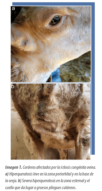
 These were characterized by multifocal hyperkeratosis, mainly affecting the muzzle, periorbital areas, and neck (Image 1).
 The affected animals showed a high fatality rate, ranging from 50% to 70%. Additionally, it was found that exposure to sunlight significantly aggravated the severity of these lesions.
Initial diagnostic approach and differential diagnosis
Based on the macroscopic lesions observed in the affected animals (Image 1) and the knowledge of ovine skin pathology (Sucupira et al., 2009), a differential diagnosis was established, which included:
 Nutritional deficiencies of zinc or vitamin A
Ectoparasites, such as fleas, mites, or lice
Superficial pyoderma
Bacterial folliculitis
Secondary photosensitization
Congenital ovine ichthyosis
In order to reach a definitive diagnosis, a clinicopathological study was conducted on a 9-month-old ewe showing signs compatible with the disease.
| The animal showed a diffuse thickening of the skin, accompanied by crusts distributed over the entire skin surface. Additionally, a lower wool density was observed, which appeared dark due to the presence of a seborrheic secretion. |
This female was mated, on the same farm, with a clinically healthy male, resulting in a twin birth.
 None of the resulting lambs showed skin lesions compatible with the disease.
Subsequently, the animal was mated again, this time with one of her sons, resulting in another twin birth.
 On this occasion, both lambs developed skin lesions, suggesting a genetic origin of the disease, with an autosomal recessive inheritance pattern.
| Based on this finding, a periodic clinical, hematological, biochemical, and histopathological follow-up was initiated to study the progression of the disease in these two affected lambs. |
Clinical and histopathological progression of the disease
Thanks to the follow-up conducted, it was possible to distinguish two phases in the progression of the disease.
1. Early phase
Appears between 3 and 5 months of age and is characterized by:

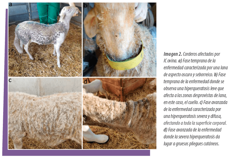
 Seborrheic, dark and clumped wool (Image 2a).
Multifocal, bilateral and symmetrical hyperkeratosis, of mild to moderate intensity, affecting wool-free areas, especially the neck, muzzle, periocular areas, and forelimbs (Image 2b)
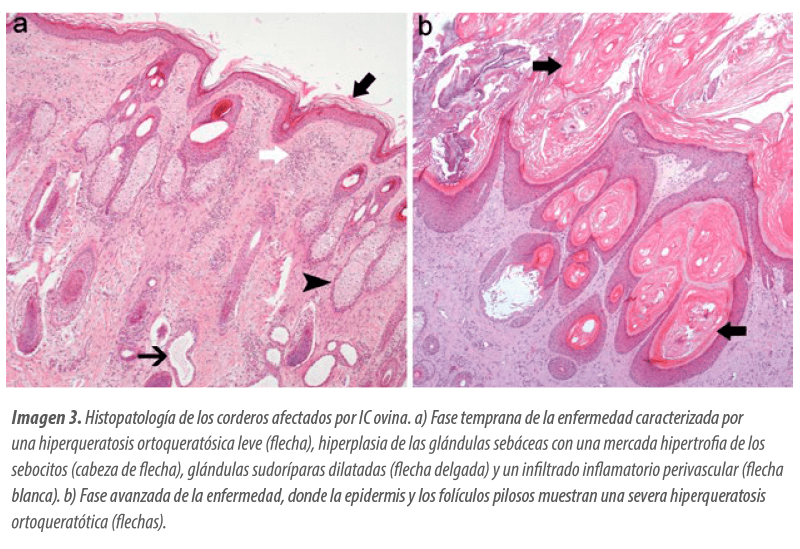
 Microscopically, this phase is defined by a laminar, diffuse and moderate orthokeratotic hyperkeratosis, accompanied by a mild perivascular, lymphoplasmacytic and eosinophilic dermatitis (Image 3a).
2. Advanced phase
It develops approximately 6 months after the appearance of the first skin lesions.
In this stage:
Hyperkeratosis extends to the wool-covered areas, affecting almost the entire body surface (Image 2c).
The skin shows markedly thickened folds, with frequent fissures and covered by thick crusts (Image 2d).
At the microscopic level, this advanced phase is characterized by:
Severe, compact, laminar orthokeratotic hyperkeratosis, arranged in concentric layers that obstruct the lumen of the hair follicles, leading in some cases to the formation of comedones (Image 3b).
Marked hyperplasia, both in the epidermis and in the follicular epithelium, while the sebaceous glands show severe atrophy.
The perivascular dermatitis also intensifies, adopting a more severe inflammatory pattern.
Diagnosis confirmation and exclusion of nutritional causes
The zinc concentrations in the feed and in the serum of the animals were found to be within the established reference values at all times (Pechin, 2017). Additionally, it was decided to administer an intramuscular injection of zinc oxide to the affected animals.
 Despite this, the skin lesions showed no improvement, which allowed a definitive exclusion of possible zinc deficiency dermatosis.
| Hematological analyses did not show any relevant alterations in the affected animals. Therefore, considering the clinical progression of the disease, the histopathological findings, and the genetic origin with an autosomal recessive inheritance pattern, the case was diagnosed as ovine IC. |
DISCUSSION: GENETIC AND CLINICAL IMPLICATIONS OF A NOVEL FINDING IN SHEEP
This study constitutes the first documented clinical case of congenital ichthyosis (CI) in the sheep species.
 The ichthyoses comprise a group of autosomal hereditary syndromes affecting the terminal differentiation of keratinocytes characterized by a localized or generalized hyperkeratosis accompanied by scaling, mainly in areas with sparse hair or wool coverage (Marukian and Choate, 2016).
 This group of diseases has been described in various animal species, including sheep, goats, cattle, swine, some canids, poultry, and even greater kudu calves.
They have been associated with mutations in multiple genes involved in various cellular functions. In veterinary medicine, two main forms of ichthyosis are recognized (Mauldin, 2016):
Lethal form or epidermolytic ichthyosis (fetalis): occurs in neonates with severe skin lesions and marked degeneration of keratinocytes (Marukian and Choate, 2016).
Non-epidermolytic or congenital form (IC): progressively affects young animals, with less severe skin lesions, compatible with life (Jacinto et al., 2021; Häfliger et al., 2022).
While ichthyosis fetalis has been previously described in sheep (Câmara et al., 2017), this study represents the first clinical and histopathological description of the congenital form in this species.
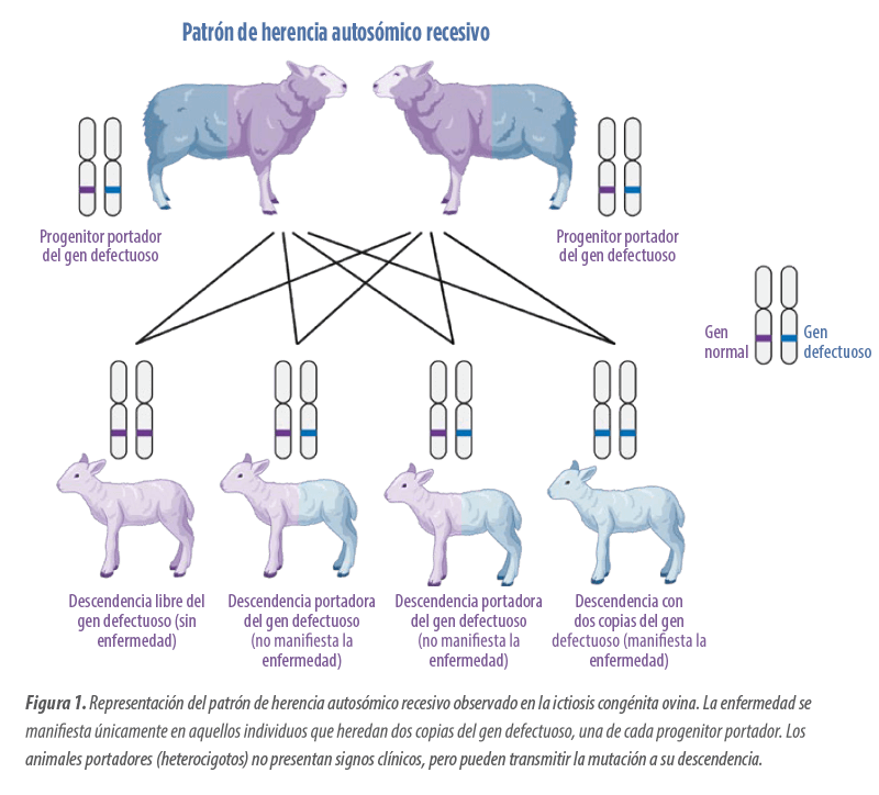
| In this disease, a clear autosomal recessive inheritance pattern is observed, which occurs when the animal inherits two defective copies of a gene (one from each parent) on a non-sex chromosome, and only then does the disease manifest. However, the gene or genes responsible for this condition in sheep have not yet been identified. |
Comparison with other species and genetic suspicion
The clinicopathological findings described in this study show similarities with bovine IC. However, in the affected lambs, skin lesions appear later (around 3 months of age) compared to cattle, where they manifest between 2 and 10 weeks of life (Raoofi, 2001; Testoni, Zappulli y Gentile, 2006; Azimpour et al., 2016; Jacinto et al., 2021; Häfliger et al., 2022).
 This suggests that the gene or genes involved in ovine IC could be different from those associated in cattle, such as FA2H and DSP (Jacinto et al., 2021; Häfliger et al., 2022).
Prevention in the herd: reproductive management keys
To prevent the occurrence of this genetic disease in sheep herds, it is essential to apply basic genetic selection measures:
 In the case of ovine IC, the reproduction of carrier animals should be avoided, recommending the removal of heterozygous individuals identified as carriers.
In the absence of specific genetic markers, the retrospective analysis of the crosses made on the farm is essential to identify the male or female carriers of the mutation.
Once removed, it is recommended to incorporate new breeders from other farms, aiming to maintain the greatest genetic diversity possible to minimize inbreeding and thereby reduce the risk of recurrence.
Diagnosis and control: an evidence-based strategy
In conclusion, the definitive diagnosis of ovine IC should be based on:
A detailed anamnesis
Histopathological analysis of the lesions
Evidence of an autosomal recessive hereditary pattern.
To rule out its main differential diagnosis, zinc deficiency dermatosis, it is essential to confirm adequate serum levels and the absence of response to supplementation with this mineral (Yuzbasiyan-Gurkan et al., 2006; Jung et al., 2014).
| The eradication of this disease in affected farms requires identifying and removing carrier animals from the breeding stock through a thorough genealogical study. |
REFERENCES
1. Azimpour, S., Sasani, F., Mottaghian, P., Anoushepour, A. 2016. Ichthyosis congenita in a calf: Case report. The Thai Journal of Veterinary Medicine, 46(3), 517–521
2. Câmara, A.C.L., Borges, P.A.C., Paiva, S.A., Pierezan, F., Soto-Blanco, B. 2017. Ichthyosis fetalis in a cross-bred lamb. Veterinary Dermatology, 28, 516-e125. https://doi.org/10.1111/vde.12459
3. Häfliger, I. M., Koch, C. T., Michel, A., Rüfenacht, S., Meylan, M., Welle, M. M., Drögemüller, C. 2022. DSP missense variant in a Scottish Highland calf with congenital ichthyosis, alopecia, acantholysis of the tongue and corneal defects. BMC Veterinary Research 18, 20. https://doi.org/10.1186/s12917-021-03113-3
4. Jacinto, J. G. P., Häfliger, I. M., Veiga, I. M. B., Letko, A., Gentile, A., Drögemüller, C. 2021. A frameshift insertion in FA2H causes a recessively inherited form of ichthyosis congenita in Chianina cattle. Molecular genetics and genomics, 296(6), 1313–1322. https://doi.org/10.1007/s00438-021-01824-8
5. Jung, S., Pausch, H., Langenmayer, M.C., Schwarzenbacher, H., Majzoub Altweck, M., Fries, R. 2014. A nonsense mutation in PLD4 is associated with a zinc deficiency-like syndrome in Fleckvieh cattle. BMC Genomics (15), 623. https://doi.org/10.1186/1471-2164-15-623
6. Marukian, N. V., Choate, K. A. 2016. Recent advances in understanding ichthyosis pathogenesis. F1000Research, 5. https://doi.org/10.12688/f1000research.8584.1
7. Mauldin, E.A., Peters-Kennedy, J. Integumentary system. In: Maxie MG, ed. Jubb, Kennedy, Palmer’s Pathology of Domestic Animals. 6th edition. St. Louis, MO: Elsevier Saunders, 2016; 509–736.
8. Pechin, G. H. 2017. Zinc in ruminant nutrition. Ciencia Veterinaria, 1 (1), 50-79
9. Raoofi, A. 2001. Ichthyosis congenita in a calf in Iran. The Veterinary record, 149(18), 563. https://doi.org/10.1136/vr.149.18.563
10. Sucupira, M. C. A., Arcaute, M. R. de Lacasta, D., Pérez, M. J., Ramos, J. J., Verde, M. T., Ferrer, L. M. 2009. Congenital and hereditary skin diseases in lambs (I). Albéitar, (128), 36-37.
11. Testoni, S., Zappulli, V., Gentile, A. (2006). Ichthyosis in two Chianina calves. Deutsche tierarztliche Wochenschrift, 113(9), 351–354.
12. Yuzbasiyan-Gurkan, V., Bartlett, E. 2006. Identification of a unique splice site variant in SLC39A4 in bovine hereditary zinc deficiency, lethal trait A46: An animal model of acrodermatitis enteropathica. Genomics, 88 (4), 521–526. https://doi.org/10.1016/j.ygeno.2006.03.018