


 Latinoamérica
 Europa
Evaluation of Two Experimental Antigens (DST-F and P22) for Immunological Diagnosis of Tuberculosis in Cattle













Bovine tuberculosis (TB) is a zoonotic mycobacteriosis caused by members of the Mycobacterium tuberculosis Complex (MTBC), especially M. bovis and M. caprae (Valcheva et al., 2022).
The presence of this disease in the livestock population entails a series of health and economic repercussions, which is why various countries, including Spain, have established programs for the eradication of TB in cattle (MAPA, 2025).
 These programs are primarily based on a diagnosis and slaughter strategy, mainly through the use of single and comparative intradermal tuberculin tests (SIT and CIT, respectively) and the interferon-gamma release assay (IGRA), combined with biosecurity and slaughterhouse surveillance.
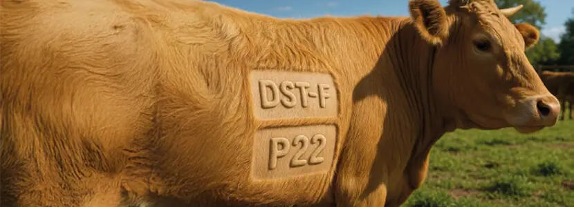
CAN THE DST-F AND P22 ANTIGENS IMPROVE THE PERFORMANCE OF BOVINE TB ANTE-MORTEM DIAGNOSTIC TESTS?
For the execution of official diagnostic tests, based on the cell-mediated immune response, antigens known as purified protein derivatives (PPDs) or tuberculins (MAPA, 2025). are used.
However, PPDs have a series of limitations:
 They present variability in their protein composition, which can influence the biological potency of the final product, with differences existing between manufacturers and even between batches due to variations in their production described in different studies (Dobbelaer et al., 1983; Good & Duignan, 2011; Balks et al., 2025).
The fact that some antigens of the PPDs are shared between CMTB mycobacteria and non-tuberculous mycobacteria, can lead to the appearance of diagnostic interferences in official tests (Fernández-Veiga et al., 2023).
For these reasons, in recent years, different lines of research have been developed with the aim of identifying, producing, and evaluating new antigens with high sensitivity and specificity, and whose production process and composition are more homogeneous and standardizable than those of PPDs, especially in the case of synthetic peptides or recombinant proteins (Middleton et al., 2021; Jones et al., 2022).
As a result of these investigations, in recent years, the following have been developed:
- Mixtures of synthetic antigens, such as ESAT-6, CFP-10, and Rv3615c (Jones et al., 2022).
- Purified immunocomplexes from bovine PPD (PPDb) such as the protein complex P22 (Infantes- Lorenzo et al., 2017).
Originally, these new antigens were mainly intended for use in in vitro techniques (IGRA and serology) (Sidders et al., 2008; Infantes-Lorenzo et al., 2019), with studies to evaluate their performance in in vivo techniques (Arrieta- Villegas et al., 2020; Jones et al., 2022) starting in recent years.
In the specific case of peptide mixtures, the initial research was aimed at obtaining a DIVA product (Differentiating Infected from Vaccinated Animals) that would allow differentiation between vaccinated and infected animals (Vordermeier et al., 2016).
However, in recent years, the lines of research concerning these antigens have evolved to diagnostic scenarios without vaccination (Kumar et al., 2021).
 The main objective was to develop alternatives to PPDs with greater specificity and similar sensitivity.
Additionally, in the case of synthetic mixtures, it would allow for the standardization of production and composition, reducing the need for biological potency tests in animals for validation, which are complex, poorly reproducible, and not always representative of the final product quality (Balks et al., 2025; Good et al., 2011).
|
Recently, studies have been conducted to evaluate the diagnostic performance of several of these antigens:
|

Use of DST-F and P22 in IDTB and IGRA
The use of these antigens in IDTB and IGRA in different species (domestic bovids and goats) has shown promising results in terms of sensitivity and specificity, so they could represent a possible alternative to PPDs in the future (Arrieta-Villegas et al., 2020; Kumar et al., 2021; Middleton et al., 2021; Jones et al., 2022; Subramanian et al., 2022).
DST-F
The DST-F has been primarily evaluated in cattle (Jones et al., 2022; Subramanian et al., 2022; Holder et al., 2023) and buffaloes (Kumar et al., 2021) in small-scale studies, generally showing a higher specificity than when using PPDb and similar sensitivity, thus constituting a potential candidate to replace it.
P22 Complex
There were no data regarding the performance of the P22 complex in relation to PPDs for conducting IDTB and IGRA in cattle.
 In goats, a small-scale study observed a better performance of both official techniques using P22 as an antigen compared to PPDs (Arrieta-Villegas et al., 2020) and has been primarily evaluated in antibody detection techniques (Ortega et al., 2022; Velasco et al., 2024).
| Large-scale field studies are needed to generate data on the performance of official diagnostic tests (IDTB and IGRA) using these two new antigens in domestic ruminants to draw conclusions about their reliability for use under different epidemiological situations in the context of TB eradication programs. |

EVALUATION OF THE PERFORMANCE OF CELL-BASED TESTS USING DST-F AND P22 AS ANTIGENS IN FIELD TESTS: THE imdiTBap PROJECT
The international project “Improving the diagnosis of tuberculosis in domestic ruminants through the use of new antigens and test platforms” (imdiTBap)” has as one of its main objectives the evaluation of the diagnostic performance of the antigens DST-F and P22 in different species of domestic ruminants and sanitary and epidemiological conditions, with the results of this article falling within it.
Study Design
Within the activities included in this project, the VISAVET center has evaluated the performance of official immunological TB diagnostic techniques in cattle using DST-F and P22 compared to the results obtained using PPDs.
 In the development of the activities necessary to achieve this objective, the performance of IDTB and IGRA in cows from four farms with TB (confirmed by bacteriological culture) was evaluated using the new antigens and traditional PPDs. In the development of the activities necessary to achieve this objective, the performance of IDTB and IGRA in cows from four farms with TB (confirmed by bacteriological culture) was evaluated using the new antigens and traditional PPDs. |
In the case of intradermal tests, the animals (n=103) were subjected to IDTBs, IDTBc, and an intradermal reaction (IDR) with DST-F (IDR-DST-F) and P22 (IDR-P22).
- Prior to performing the intradermal tests, blood samples were taken from these same animals for subsequent stimulation with traditional PPDs, DST-F, and P22 and to perform the IGRA technique with the commercial Bovigam kit (Thermo Fisher Scientific, Waltham, USA).
- The interpretation criteria for the different intradermal tests (IDTBs, IDTBc, and IDR) and the IGRA technique are described in the Tables 1 and 2.
 To establish the differences regarding the proportion of reactors to the different techniques using different antigens in the study, they were statistically analyzed using the Cochran’s Q test and McNemar’s test, adjusting the p-value with the Bonferroni correction and considering a p value of 0.05 as statistically significant.

Preliminary results
 The highest number of reactors to the intradermal tests (IDTBs-IDR) was detected when using P22 as the antigen, both using a standard interpretation criterion (7/103; 6.8%) and severe (10/103; 9.7%), followed by PPDb (6/103; 5.8% and 8/103; 7.8%; respectively) and DST-F (4/103; 3.9% with both interpretations).
In the case of DST-F, the number of reactors increased using an extra-severe cutoff point (recommended by the manufacturer) obtaining a number of reactors similar to that observed with the standard interpretation of IDR-P22 (7/103; 6.8%) and slightly higher than that of IDTBs (6/103; 5.8%).
In contrast, the use of standard and severe interpretations (4/103; 3.9%) of IDR-DST-F led to a reactivity of the technique similar to IDTBc, especially to its severe interpretation (4/103; 3.9%) (Table 1 and Graph 1).
 There were significant differences in the proportion of reactors when using the different antigens (p < 0.05):
 The number of reactors to the severe interpretation of the IDR-P22 was significantly higher (p < 0.05) compared to that observed with the same interpretation of the IDR-DST-F (Figure 1).
A higher number of reactors was observed when using P22 as an antigen compared to PPDb, contrary to what was observed in a previous study in goats (Arrieta-Villegas et al., 2020).
| A possible explanation could be the differences between species, with this greater reactivity of intradermal tests using P22 as an antigen being characteristic of the bovine species. |


Regarding DST-F, the lower reactivity shown with this antigen in intradermal tests compared to PPDb has been observed in previous studies in infected cattle (Jones et al., 2022; Holder et al., 2023).
However, further studies are needed to confirm the findings observed in this study, especially regarding the differences observed in reactivity between P22 and DST-F, as this is the first study comparing both antigens in this species.
Regarding the performance shown by the IGRA, the results of the reactivity obtained are described in the Table 2.

Very similar reactivities were obtained with the three antigens (PPDb, DST-F, and P22) using the Bovigam kit (Table 2 and Figure 2).
In fact, no significant differences (p > 0.05) were observed in the stimulated samples (n=103) with the different antigens that resulted positive, obtaining a percentage of reactors of 20.4% (21/103), 21.4% (22/103), and 22.3% (23/103) using PPDb, DST-F, and P22, respectively.

| The higher reactivity shown with P22 is consistent with what was previously observed in goats with this same kit (Arrieta-Villegas et al., 2020). Considering the results obtained by stimulating the samples with DST-F, the reactivity achieved with this antigen, similar to that observed with PPDb, has been previously described in this species (Holder et al., 2023). |

CONCLUSIONS
In the present study, the highest diagnostic performance of the intradermal tests (IDTB/IDR) was obtained using P22 as an antigen, followed by PPDb and DST-F, the latter being very similar to that observed when applying IDTBc and significantly lower than that observed with P22.
In contrast, no significant differences were observed in the diagnostic performance shown by the IGRA using PPDb, DST-F and P22, with minimal variations in the reactivity shown by the different antigens.
| These preliminary results provide valuable information on the usefulness of DST-F and P22 as antigens in official immunological-based diagnostic tests for TB in cattle, with a need to confirm these findings with results in free herds and analyses conducted by other countries participating in the imdiTBap project. |
Acknowledgments
This research has been made possible thanks to funding from ICRAD, an ERA-NET network co-funded by the European Union’s Horizon 2020 research and innovation program under grant agreement nº862605, and the Ministry of Science, Innovation and Universities of Spain (MCIN/AEI/10.13039/501100011033) through the project “Improving the diagnosis of tuberculosis in domestic ruminants through the use of new antigens and test platforms” (reference PCI2023-143368), and the Ministry of Agriculture, Fisheries and Food of Spain.

BIBLIOGRAPHY
1. Arrieta-Villegas, C., Infantes-Lorenzo, J. A., Bezos, J., Grasa, M., Vidal, E., Mercader, I., Singh, M., Domingo, M., de Juan, L., & Pérez de Val, B. (2020). Evaluation of P22 Antigenic Complex for the Immuno-Diagnosis of Tuberculosis in BCG Vaccinated and Unvaccinated Goats. Frontiers in veterinary science, 7, 374. https://doi.org/10.3389/fvets.2020.00374
2. Balks, E., Trösemeier, J. H., Mühlebach, M. D., & Reuter, A. (2025). Mass spectrometry analysis of bovine tuberculins – 3R potential in batch potency testing. Biologicals: journal of the International Association of Biological Standardization, 90, 101825. https://doi.org/10.1016/j.biologicals.2025.101825
3. Dobbelaer, R., O’Reilly, L. M., Génicot, A., & Haagsma, J. (1983). The potency of bovine PPD tuberculins in guinea-pigs and in tuberculous cattle. Journal of biological standardization, 11(3), 213–220. https://doi.org/10.1016/s0092-1157(83)80008-0
4. Fernández-Veiga, L., Fuertes, M., Geijo, M. V., Pérez de Val, B., Vidal, E., Michelet, L., Boschiroli, M. L., Gómez-Buendía, A., Bezos, J., Jones, G. J., Vordermeier, M., Juste, R. A., Garrido, J. M., & Sevilla, I. A. (2023). Differences in skin test reactions to official and defined antigens in guinea pigs exposed to non-tuberculous and tuberculous bacteria. Scientific reports, 13(1), 2936. https://doi.org/10.1038/s41598-023-30147-4
5. Good, M., Clegg, T. A., Murphy, F., & More, S. J. (2011). The comparative performance of the single intradermal comparative tuberculin test in Irish cattle, using tuberculin PPD combinations from different manufacturers. Veterinary microbiology, 151(1-2), 77–84. https://doi.org/10.1016/j.vetmic.2011.02.028
6. Good, M., & Duignan, A. (2011). Perspectives on the History of Bovine TB and the Role of Tuberculin in Bovine TB Eradication. Veterinary medicine international, 2011, 410470. https://doi.org/10.4061/2011/410470
7. Holder, T., Coad, M., Allan, G., Hogarth, P. J., Vordermeier, H. M., & Jones, G. J. (2023). Vaccination of calves with Bacillus Calmette-Guerin Danish strain 1331 results in a duration of immunity of at least 52 weeks. Vaccine, 41(48), 7290–7296. https://doi.org/10.1016/j.vaccine.2023.10.060
8. Infantes-Lorenzo, J. A., Moreno, I., Risalde, M. L. Á., Roy, Á., Villar, M., Romero, B., Ibarrola, N., de la Fuente, J., Puentes, E., de Juan, L., Gortázar, C., Bezos, J., Domínguez, L., & Domínguez, M. (2017). Proteomic characterisation of bovine and avian purified protein derivatives and identification of specific antigens for serodiagnosis of bovine tuberculosis. Clinical proteomics, 14, 36. https://doi.org/10.1186/s12014-017-9171-z
9. Infantes-Lorenzo, J. A., Moreno, I., Roy, A., Risalde, M. A., Balseiro, A., de Juan, L., Romero, B., Bezos, J., Puentes, E., Åkerstedt, J., Tessema, G. T., Gortázar, C., Domínguez, L., & Domínguez, M. (2019). Specificity of serological test for detection of tuberculosis in cattle, goats, sheep and pigs under different epidemiological situations. BMC veterinary research, 15(1), 70. https://doi.org/10.1186/s12917-019-1814-z
10. Jones, G. J., Konold, T., Hurley, S., Holder, T., Steinbach, S., Coad, M., Neil Wedlock, D., Buddle, B. M., Singh, M., & Martin Vordermeier, H. (2022). Test performance data demonstrates utility of a cattle DIVA skin test reagent (DST-F) compatible with BCG vaccination. Scientific reports, 12(1), 12052. https://doi.org/10.1038/s41598-022-16092-8
11. Köhler, H., Liebler-Tenorio, E., Hughes, V., Stevenson, K., Bakker, D., Willemsen, P., Bay, S., Ganneau, C., Biet, F., & Vordermeier, H. M. (2021). Interferon-γ Response of Mycobacterium avium subsp. paratuberculosis Infected Goats to Recombinant and Synthetic Mycobacterial Antigens. Frontiers in Veterinary Science, 8, 645251. https://doi.org/10.3389/fvets.2021.645251
12. Kumar, T., Singh, M., Jangir, B. L., Arora, D., Srinivasan, S., Bidhan, D., Yadav, D. C., Veerasami, M., Bakker, D., Kapur, V., & Jindal, N. (2021). A Defined Antigen Skin Test for Diagnosis of Bovine Tuberculosis in Domestic Water Buffaloes (Bubalus bubalis). Frontiers in veterinary science, 8, 669898. https://doi.org/10.3389/fvets.2021.669898
13. MAPA. Ministerio de Agricultura Pesca y Alimentación. Programa Nacional de Erradicación de Tuberculosis Bovina 2025 (Infección por el complejo Mycobacterium tuberculosis). (2025). Available online at: https://www.mapa.gob.es/es/ganaderia/temas/sanidad-animal-higiene-ganadera/programatb2025_18022025_tcm30-698268.pdf
14. Middleton, S., Steinbach, S., Coad, M., McGill, K., Brady, C., Duignan, A., Wiseman, J., Gormley, E., Jones, G. J., & Vordermeier, H. M. (2021). A molecularly defined skin test reagent for the diagnosis of bovine tuberculosis compatible with vaccination against Johne’s Disease. Scientific reports, 11(1), 2929. https://doi.org/10.1038/s41598-021-82434-7
15. Ortega, J., Infantes-Lorenzo, J. A., Roy, Á., de Juan, L., Romero, B., Moreno, I., Domínguez, M., Domínguez, L. & Bezos, J. (2022). Factors affecting the performance of P22 ELISA for the diagnosis of caprine tuberculosis in milk samples. Research in Veterinary Science, 145, 40–45. https://doi.org/10.1016/j.rvsc.2022.02.008
16. Sidders, B., Pirson, C., Hogarth, P. J., Hewinson, R. G., Stoker, N. G., Vordermeier, H. M., & Ewer, K. (2008). Screening of highly expressed mycobacterial genes identifies Rv3615c as a useful differential diagnostic antigen for the Mycobacterium tuberculosis complex. Infection and immunity, 76(9), 3932–3939. https://doi.org/10.1128/IAI.00150-08
17. Subramanian, S., Srinivasan, S., Ramaiyan Selvaraju, K., Vinoli, P. M., Selladurai, S., Ramasamy, B., Kumaragurubaran, K., Bakker, D., Vordermeier, M., Kapur, V., & Gopal, D. R. (2022). Defined Antigen Skin Test for Bovine Tuberculosis Retains Specificity on Revaccination With Bacillus Calmette-Guerin. Frontiers in Veterinary Science, 9, 814227. https://doi.org/10.3389/fvets.2022.814227
18. Valcheva, V., Perea, C., Savova-Lalkovska, T., Dimitrova, A., Radulski, L., Mokrousov, I., Marinov, K., Najdenski, H., & Bonovska, M. (2022). Mycobacterium bovis and M. caprae in Bulgaria: insight into transmission and phylogeography gained through whole-genome sequencing. BMC Veterinary Research, 18(1), 148. https://doi.org/10.1186/s12917-022-03249-w
19. Velasco, C., Ortega, J., Ricón, J., Romero, B., de Juan, L., Domínguez, L., Domínguez, M., Moreno, I., Álvarez, J., & Bezos, J. (2024). Effect of a recent intradermal test on the specificity of P22 ELISA for the diagnosis of caprine tuberculosis. Frontiers in veterinary science, 11, 1358413. https://doi.org/10.3389/fvets.2024.1358413
20. Vordermeier, H. M., Jones, G. J., Buddle, B. M., & Hewinson, R. G. (2016). Development of immune-diagnostic reagents to diagnose bovine tuberculosis in cattle. Veterinary immunology and immunopathology, 181, 10–14. https://doi.org/10.1016/j.vetimm.2016.02.003