


 Latinoamérica
 Europa
Exploration of the Respiratory System Using Ultrasound in Sheep


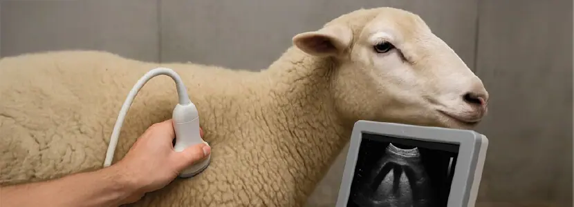










The lung ultrasound is of growing interest in the livestock sector. In fact, in cattle, its use is already widespread in some phases of the production cycle, such as, for example, in the rearing of dairy heifers or in the incorporation into feedlots of calves.
 Now, this technique is also starting to gain relevance in sheep, a species in which respiratory diseases have a significant health and productive impact.

In the case of sheep:
 The lung of the sheep has good capacity to transmit ultrasound, which allows the use of intermediate frequencies between 5.0 and 6.5 MHz in adults.
In young lambs or when exploring more superficial structures, it may be useful to use higher frequencies to improve image resolution (up to 10 MHz).
 For this type of examination, convex or microconvex probes can be used, as their curved shape and trapezoidal image facilitate access between the ribs and offer good visualization. The linear probe for larger sheep is also valid. For this type of examination, convex or microconvex probes can be used, as their curved shape and trapezoidal image facilitate access between the ribs and offer good visualization. The linear probe for larger sheep is also valid. |
Technique and positioning in thoracic exploration
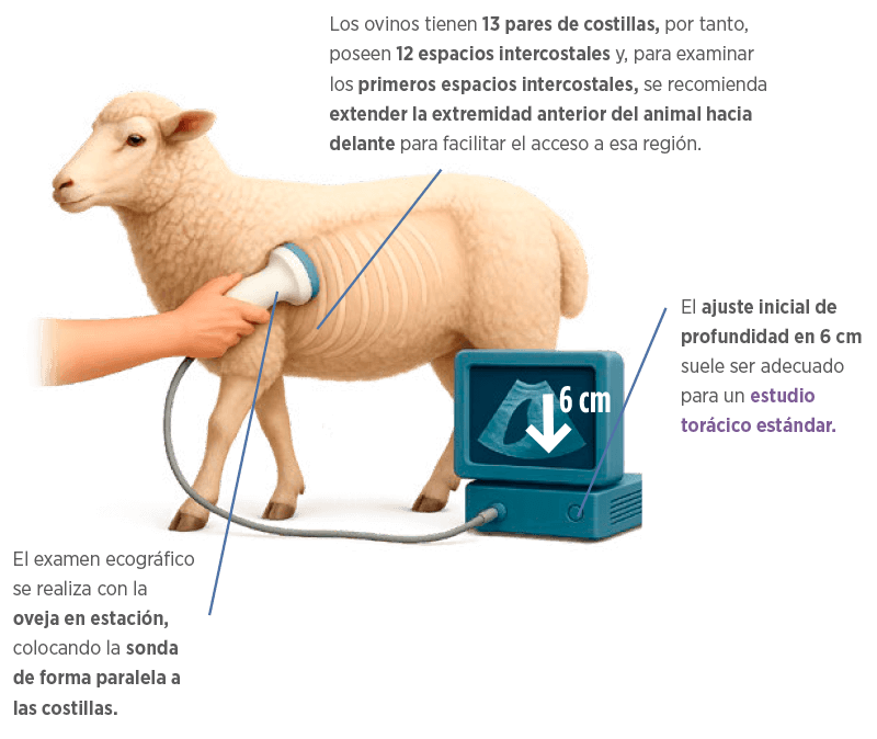
| It is important to examine both sides of the thorax, as lesions can be unilateral or vary in severity. |
Ultrasound patterns in healthy and injured lungs
HEALTHY LUNG 
 In a healthy lung, a continuous hyperechoic line is observed in the upper region, corresponding to the visceral pleura, which represents the interface with the lung surface.
Being a predominantly aerated organ, the lung generates reverberation artifacts known as A-lines, which appear as parallel horizontal lines and are a normal ultrasound finding.
This image may vary depending on the age or body condition of the animal.
ALTERED LUNG
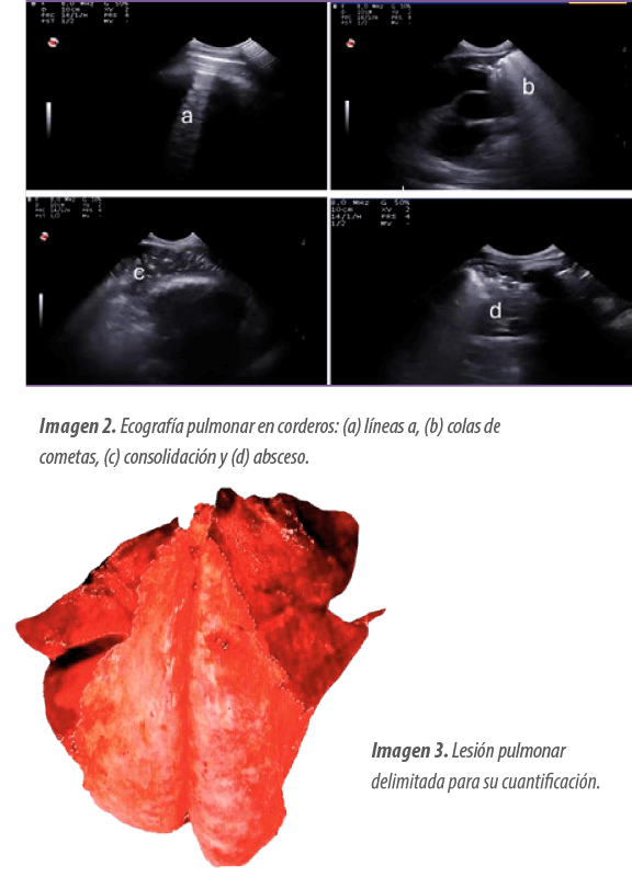
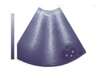 Among the most common alterations are B-lines or “comet tails”, which indicate inflammation or fluid in the tissue.
Pleural irregularities, thickenings, or discontinuities can be observed, suggestive of infection, as well as pulmonary consolidations, where the tissue loses its airy pattern and becomes denser.
Pulmonary consolidations can be localized or extensive and, in severe cases, can give a liver-like appearance (hepatization), characteristic of advanced pneumonias.
Within these areas, it is common to visualize an air bronchogram, represented by bronchi filled with air as hyperechoic dots or lines.
The most frequently observed lower respiratory tract diseases in adult sheep include:
- Ovine Respiratory Complex (ORC).
- Ovine Pulmonary Adenocarcinoma (OPA).
- Aspiration pneumonia (gangrenous).
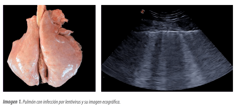
- Pulmonary form of small ruminant lentivirus infection (SRLV, Image 1).
- Visceral form of caseous lymphadenitis.
- Verminous pneumonia caused by parasitic lungworms.
 All of these are potentially detectable by lung ultrasound.
| Certain lung diseases may be more commonly associated with specific areas, so it is also important to relate the different images to the region being evaluated (Table 1). |
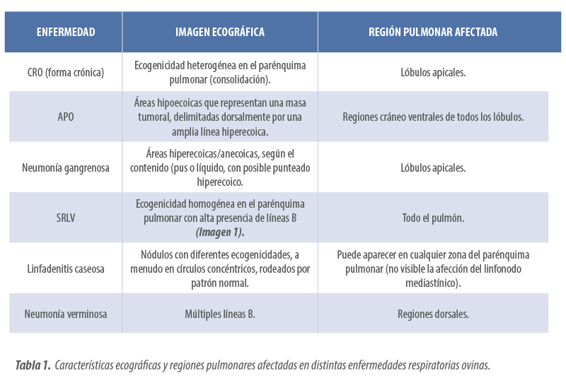
| It is important to note that the simultaneous occurrence of multiple lung diseases in the same adult individual is a very common phenomenon, so it should be considered during the ultrasound evaluation of the lung. |

LUNG ULTRASOUND IN LAMBS
 The application of lung ultrasound in adult sheep has a promising future, already being a standardized technique in the United Kingdom for the diagnosis of OPA.
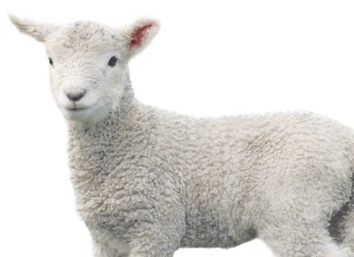
In contrast, in the case of lambs, the most plausible application would be the diagnosis of PRD, which is one of the main causes of morbidity and mortality during the fattening phase, severely affecting animal welfare and the profitability of livestock farms.
|
The early and accurate detection of lung lesions is essential for:
|
 Optimizing treatment.
Field diagnostic evaluation
 One of the most recent studies by this team aimed to evaluate the utility of thoracic ultrasound in lambs through comparison with conventional diagnostic methods such as auscultation, clinical scoring, and postmortem evaluation.
This study was conducted on a dairy sheep farm of the Lacaune breed located in the Valencian Community (Spain), with a sample of 111 lambs.
After weaning and during the fattening phase, the animals were selected for showing clinical signs or conditions likely to delay growth, being suitable for transport to the slaughterhouse.
Diagnostic tests were performed 12 hours before slaughter.
|
|
 The lung auscultation was carried out using a systematized protocol with a stethoscope, evaluating the right and left lung lobes described as:
- Normal sounds.
- Bronchial sounds.
- Snoring.
- Crackling sounds.
- Absence of respiratory sounds.
|
|
 Thoracic ultrasound was performed with a portable ultrasound machine (Esaote MyLab One Vet) equipped with a 10 MHz microconvex probe. In this, the cranial, middle, and caudal lung lobes were sequentially evaluated, categorizing the obtained images according to the appearance of:
 In the slaughterhouse, the postmortem evaluation was performed, considered as the reference technique, measuring the percentage of affected lung and classifying the macroscopic lesions in a standardized manner into four categories (Image 3).
| The severity and extent of the disease, assessed according to each of the four techniques (physical examination, auscultation, pulmonary ultrasound, and postmortem evaluation) was rated from 0 to 3. Sensitivity and correlation between the techniques were then calculated, using macroscopic postmortem evaluation as a reference. |
Limitations of conventional techniques
Despite being a cost-effective and quick technique, auscultation has limited diagnostic utility compared to ultrasound due to its low specificity, difficulty in locating abscesses or distinguishing lung sounds from other physiological sounds, and also due to the operator’s influence.
Although useful in moderate cases, its ability to assess the severity of processes and correlation with postmortem evaluation was lower.
 The physical examination is a valuable tool for initial screening and decision-making in the field, but it may not detect the mildest or incipient cases as it does not detect subclinical cases (24.3% of animals that did not show clinical signs exhibited ultrasound alterations compatible with respiratory disease).
Furthermore, it does not have sufficient sensitivity to classify cases according to their severity.
Advantages of lung ultrasound in lambs
The lung ultrasonography allowed for an objective, non-invasive, and repeatable evaluation of lung lesions, with the ability to identify consolidations, B-lines, effusions, and abscesses, even in animals without clinical symptoms.
Although it presents limitations in deep or mediastinal lesions, its application in lambs during the fattening phase is particularly useful, given the superficial location and the typical location of pneumonias in the cranioventral regions of the thorax. Additionally, it can serve to monitor the response to treatment and to implement preventive strategies based on management or vaccination.
| In conclusion, this study demonstrates that lung ultrasound is a reliable, practical, and complementary tool for the diagnosis of the ovine respiratory complex.
|
 Its incorporation into routine diagnostic protocols is recommended, as well as conducting future studies to relate ultrasound patterns with specific etiological agents. Its incorporation into routine diagnostic protocols is recommended, as well as conducting future studies to relate ultrasound patterns with specific etiological agents. |

You may be interested in: Multicentric lymphoma in a sheep