


 Latinoamérica
 Europa
Squamous Cell Tumor in a Culling Sheep














Squamous cell carcinoma is a malignant neoplasm of epithelial origin that, although common in domestic species such as cattle or horses, occurs more sporadically in sheep.
This article describes a clinical case of periocular squamous cell carcinoma in a sheep, detailing the clinical, diagnostic, and pathological findings, as well as considerations for its management and prevention under field conditions.
AN EYE LESION WITH AN UNEXPECTED DIAGNOSIS
Background and reason for consultation 
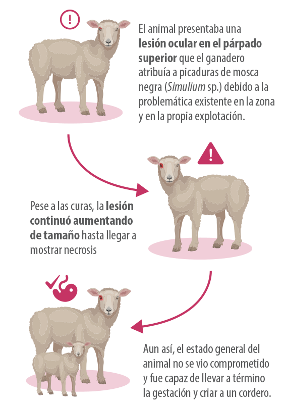
A 7-year-old Rasa Aragonesa sheep from a farm located in the Ribera del Ebro was referred to the Ruminant Clinical Service (SCRUM) of the University of Zaragoza.
Clinical examination: a mass that kept advancing
After the weaning of the lamb, the animal was referred to the SCRUM of the University of Zaragoza, where a thorough clinical examination was performed.
 The examination revealed a body condition score of 2/5 and the presence of serous nasal discharge, with no abnormalities upon auscultation.
The most relevant finding was the mass located at the facial level, specifically on the right lower eyelid, causing protrusion and deformation of the periocular tissues, with occlusion of the medial canthus and lateral deviation of the eyeball within the orbit.
 The tumor surface was crusted and ulcerated, with the presence of a serosanguineous discharge (Figure 1).

Complementary tests: what the data showed
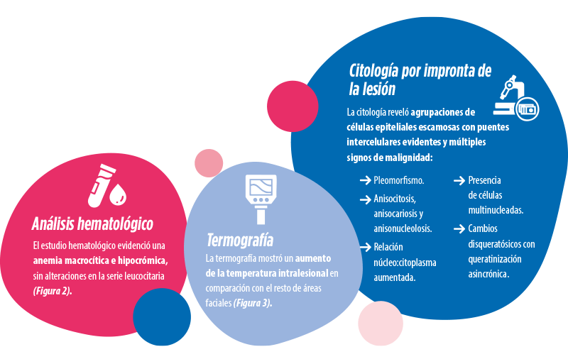
The following complementary tests were performed:


From differential diagnosis to definitive diagnosis
Initially, in the differential diagnosis of the condition presented by the animal, various pathologies were considered, including:
 Myiasis by fly larvae (families Calliphoridae and Sarcophagidae).
Infected lesions after blackfly bites.
Oestrosis caused by Oestrus ovis larvae.
Traumatic causes.
Periocular abscesses.
Neoplastic processes, such as enzootic nasal adenocarcinoma (ENA), characterized by lesions in the ethmoid turbinates and nasal discharge, as well as other cutaneous or ocular tumors, including melanoma, fibroma, fibrosarcoma, and squamous cell carcinoma.
Subsequently, due to the compromise that the lesion posed to the animal’s welfare and its progressive worsening, it was referred to the Veterinary Anatomopathological Diagnostic Service of the University of Zaragoza for necropsy and macroscopic examination of the lesions.
 Regarding the periocular lesion (Figure 4), the removal of the skin revealed a multinodular mass infiltrating the superficial soft tissues (skin and conjunctiva), without bone or ocular invasion. Partial sectioning of the mass showed extensive areas of necrosis. Regarding the periocular lesion (Figure 4), the removal of the skin revealed a multinodular mass infiltrating the superficial soft tissues (skin and conjunctiva), without bone or ocular invasion. Partial sectioning of the mass showed extensive areas of necrosis. |

Among the additional post-mortem findings were observed:
Abdominal cysticercosis along with mild hepatic degeneration and ruminal parakeratosis.
Pleuritic adhesions in the thoracic cavity, compatible with fibrous pleuritis.
Multifocal granulomatous bronchopneumonia in the diaphragmatic lobes, compatible with verminous pneumonia caused by protostrongylid nematodes.
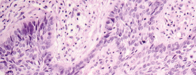
The histopathological study of the periocular mass showed numerous signs of malignancy:
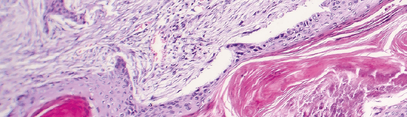
 The mass was composed of cords and islands of neoplastic squamous cells supported by an abundant fibrovascular stroma.
The cords presented central areas of keratinization with concentric laminar keratin formation in the form of horny pearls
(Figure 5).
At higher magnifications, the neoplastic population showed a marked cellular and nuclear pleomorphism, with prominent nucleoli and karyomegaly (Figure 6).
The fibrovascular stroma was infiltrated by abundant leukocytes, lymphocytes and macrophages, with frequent hemorrhages (Figure 6).


 At the microscopic level, in the lung, a peribronchial inflammatory infiltrate mainly composed of lymphocytes was observed. The liver showed changes compatible with fatty degeneration or hepatic steatosis. At the microscopic level, in the lung, a peribronchial inflammatory infiltrate mainly composed of lymphocytes was observed. The liver showed changes compatible with fatty degeneration or hepatic steatosis. |
DISCUSSION
Diagnostic confirmation and clinical relevance
The persistence of the lesion and the set of clinicopathological findings allowed for the exclusion of most differential diagnosis options and directed the case towards a neoplasm.
The histopathological study confirmed the diagnosis of squamous cell carcinoma, a malignant neoplasm, locally infiltrative and rapidly growing.
 This type of tumor, uncommon in the sheep species, appears sporadically and has been described in various countries with high insolation such as Uruguay, Brazil, Australia, Iran, Italy, or Spain (Costa et al., 2019; Ladds et al., 1977; Méndez et al., 1997; Ramos et al., 2007).
 The consulted references emphasize that the most valuable diagnostic tools are cytology and, especially, histopathology. The consulted references emphasize that the most valuable diagnostic tools are cytology and, especially, histopathology. |

Predisposing factors and mechanisms involved
The main predisposing factor is exposure to solar radiation (García et al., 2018), although other factors may also be involved, such as infection by papillomavirus (Vitiello et al., 2017), trauma (García et al., 2018) or a possible genetic predisposition (Vasconcelos et al., 2023).
 Susceptibility is higher in adult animals and, given the suggested heritability component, genetic selection of individuals could be a preventive tool (Vasconcelos et al., 2023).
 The influence of UV radiation on the occurrence of the tumor makes it advisable to limit sun exposure through appropriate management measures. The influence of UV radiation on the occurrence of the tumor makes it advisable to limit sun exposure through appropriate management measures. |
In pregnant females, as in the case described here, it is essential to take extreme care, as factors such as nutritional deficiencies, increased stress, or immunosuppression could promote the development of this type of carcinomas (Becerra and Ruano, 2015).
In fact, the presented case showed underlying hematological alterations, such as macrocytic and hypochromic anemia.

Therapeutic limitations and preventive approach
Currently, there are no established therapeutic protocols for sheep.
In other domestic species, such as companion animals, surgery is considered the treatment of choice. However, in production animals, the complete surgical resection of periocular masses is usually not feasible.
This limitation is compounded by the high presence of flies and other insects in this region of Spain for much of the year, which complicates postoperative management and compromises proper wound healing.
All this reinforces the need to emphasize preventive measures and the reduction of predisposing factors.
CONCLUSION
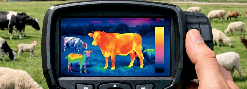
The diagnosis of periocular squamous cell carcinoma in sheep is based on anamnesis and clinical examination, complemented by tests such as imprint cytology, hematology, and thermography.
The definitive diagnosis depends on the pathological study of the proliferative tissue, which must show characteristic malignancy criteria, such as pleomorphism, infiltrative pattern, and/or presence of keratin pearls, among others.
Squamous cell carcinoma should be included among the differential diagnoses of ocular and periocular masses in sheep, along with other tumors described in ruminants.
Among the possible etiological factors are ultraviolet (UV) radiation, genetic factors, and even viral factors in some cases, so its management must be fundamentally preventive.
BIBLIOGRAPHY
University of Murcia. (2012). Neoplasia – Cytology. Innova OCW. https://www.um.es/innova/OCW/PropedeuticaClinica2012/material_clase/Citologia/neoplasia.html
Becerra Pérez, D. (2016). Squamous cell carcinoma (Case report). Available at: https://repository.udca.edu.co/handle/11158/504
Costa, R. A., Schild, C., Caffarena, D., Giannitti, F., Banchero, G., Del Pino, M. L., & Riet‑Correa, F. (2019). High frequency of cutaneous squamous cell carcinoma in Friesian Milchschaf sheep in Uruguay. Pesquisa Veterinária Brasileira, 39(4), 251–254. https://doi.org/10.1590/1678-5150-PVB-6170
De la Cruz-Cruz, L., Pérez, E., Larrondo Cornejo, C., García, J., & Roldán-Santiago, P. (2022). Use of infrared thermography in the detection of health problems in ruminants. Uso de la termografía infrarroja en la detección oportuna de problemas de salud en rumiantes.
Ferrer, L., Lacasta, D., Ramos, J., Jalón, J., Ruiz De Arcaute, M., & Conde, T. (2011). Squamous cell carcinoma of the vagina and cervix in sheep — Case report. Acta Veterinaria Hungarica, 59(1), 123–127
García, J. A., Quinteros, C., Romero, A., & Dutra, F. (2018). Occurrence of squamous cell carcinoma in Milchschaf sheep in Uruguay. Ciência Rural, 48(1), e20170406. https://doi.org/10.1590/0103-8478cr20170406
Ladds, P. W., & Entwistle, K. W. (1977). Observations on squamous cell carcinomas of sheep in Queensland, Australia. British journal of cancer, 35(1), 110–114. https://doi.org/10.1038/bjc.1977.10
Méndez, A., Abella, A., & López, A. (1997). Clinicopathological study of an outbreak of squamous cell carcinoma in sheep in south-west Spain. Veterinary Record, 140(26), 688-690. https://doi.org/10.1136/vr.140.26.688
Ramos, A. T., Norte, D. M., Elias, F., & Fernandes, C. G. (2007). Squamous cell carcinoma in cattle, sheep and horse: Study of 50 cases in south of Rio Grande do Sul. Brazilian Journal of Veterinary Research and Animal Science, 44(supl.), 5–13. https://doi.org/10.11606/issn.1678-4456.bjvras.2007.26583
Vasconcelos, J., Pires, M. d. A., Alves, A., Vieira-Pinto, M., Saraiva, C., & Cardoso, L. (2023). Neoplasms in domestic ruminants and swine: A systematic literature review. Veterinary Sciences, 10(2), 163. https://doi.org/10.3390/vetsci10020163
Vitiello, V., Burrai, G. P., Agus, M., Anfossi, A. G., Alberti, A., Antuofermo, E., Rocca, S., Cubeddu, T., & Pirino, S. (2017). Ovis aries papillomavirus 3 in ovine cutaneous squamous cell carcinoma. Veterinary Pathology, 54(5), 775–782. https://doi.org/10.1177/0300985817705171